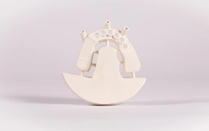
An implant surgical guide is a patient-specific template that supports the accurate placement of dental implants during surgery. It is designed using digital planning software based on CBCT data and intraoral or laboratory scans, then manufactured using CAD/CAM technology.
The guide fits precisely onto the patient's teeth, mucosa or bone and contains guide sleeves that direct the surgical drills according to the virtual implant plan.
Because the guide transfers the digital planning directly to the clinical procedure, it minimizes deviations between the planned and the final implant position.
Modern surgical guides contribute to:
For clinicians restoring complex implant cases, guided surgery has become an important tool for achieving predictable outcomes.
How does a surgical guide work?
The fabrication process begins with digital patient data. A cone beam computed tomography (CBCT) scan provides detailed information about the patient's bone anatomy, while an intraoral scan captures the soft tissue and remaining dentition.
These datasets are combined within implant planning software to create a comprehensive three-dimensional representation of the patient's anatomy.
After the implant position has been planned virtually, the software generates a customized guide that fits the patient's anatomy precisely. Guide sleeves incorporated into the design ensure that the drills follow the planned angulation and depth during surgery.
This digital workflow significantly improves the transfer of treatment planning into clinical reality.

Not every implant case requires the same type of guide. The supporting anatomy and clinical indication determine which guide design is most appropriate.
The three most common guide types are tooth-supported, mucosa-supported and bone-supported guides.
Guide Type | Support | Typical Indications |
|---|---|---|
| Tooth-supported | Remaining natural teeth | Single implants, short-span restorations, partially edentulous patients |
| Mucosa-supported | Soft tissue | Fully edentulous patients, flapless surgery |
| Bone-supported | Alveolar bone | Extensive bone augmentation, complex implant surgery |
Tooth-supported guides
Tooth-supported guides are considered the most stable and accurate option whenever sufficient natural teeth are present.
The guide rests on the remaining dentition, providing excellent positional stability throughout the surgical procedure.
These guides are commonly used for:
Because the support is provided by rigid tooth structures, tooth-supported guides generally achieve the highest level of accuracy.
Mucosa-supported guides
For completely edentulous patients, tooth support is no longer available.
In these cases, the guide is designed to rest directly on the patient's mucosa.
Mucosa-supported guides are frequently used for:
To improve stability, fixation pins are often incorporated into the guide design.
Although mucosal support introduces slightly greater movement potential than tooth-supported guides, careful planning and fixation allow highly predictable clinical outcomes.
Bone-supported guides
Bone-supported guides are primarily reserved for complex surgical cases.
After reflecting the soft tissue, the guide is positioned directly on the exposed alveolar bone.
Typical indications include:
Because these guides require flap elevation, they are generally used for more complex surgical procedures rather than routine implant placement.
Selecting the appropriate guide type is one of the first and most important decisions during digital implant planning, as it influences guide stability, surgical workflow and overall treatment predictability.

Modern CAD/CAM software allows clinicians and dental technicians to design highly accurate implant surgical guides using a fully digital workflow.
The process begins long before manufacturing. Every successful guide depends on precise digital data, careful implant planning and a well-designed guide geometry.
Step 1: Import CBCT data
The workflow starts with importing the patient's CBCT scan into icam.
CBCT data provides a three-dimensional representation of the patient's anatomical structures, including:
The DICOM data generated by the CBCT scanner is imported into the software and forms the basis of implant planning.
To create an accurate restorative workflow, the CBCT dataset is then aligned with the digital surface scan obtained from an intraoral scanner or laboratory scanner.
This matching process combines hard tissue information with highly detailed surface anatomy, creating a complete virtual patient model.
Accurate alignment of these datasets is essential, as even minor registration errors can influence the final implant position.
Step 2: Implant planning in icam
After the datasets have been merged successfully, implant planning begins.
Rather than positioning implants solely according to available bone, modern digital workflows follow a prosthetically driven approach. This means the desired final restoration determines the ideal implant position, while anatomical limitations such as bone volume, neighbouring teeth and vital structures are carefully considered.
Within icam, users can virtually position implants while evaluating:
The software enables clinicians to inspect the implant position simultaneously in axial, sagittal, coronal and three-dimensional views, making it easier to assess the relationship between the implant and the surrounding anatomy.
This comprehensive visualization helps reduce the risk of surgical complications while supporting prosthetically ideal implant placement.
Step 3: Guide design
Once the implant positions have been finalized, the surgical guide itself can be designed.
Based on the selected guide type, icam automatically generates the basic guide geometry, which can then be refined according to the clinical requirements.
During this stage, the user defines important design parameters such as guide thickness, extension and retention areas to ensure a stable fit during surgery.
Particular attention is given to the positioning of the guide sleeves. These sleeves determine the drilling path and therefore directly influence surgical accuracy.
In addition, the software allows the designer to optimize support surfaces, improve insertion paths and adapt the guide to the patient's anatomy while maintaining sufficient mechanical stability.
A carefully designed surgical guide not only improves manufacturing reliability but also contributes to easier handling during the surgical procedure.
Step 4: Milling the surgical guide with coritec
Once the guide design has been finalized, the CAD model is transferred to the CAM environment, where the machining strategy is generated. Based on the selected material and the geometry of the guide, the software calculates the appropriate toolpaths before sending the manufacturing data to the coritec milling machine.
Surgical guides are typically milled from transparent polymer discs that offer excellent dimensional stability and visibility during surgery. The high precision of modern milling systems ensures that critical features such as guide sleeves, support surfaces and fitting geometry are reproduced accurately.
Compared with manual fabrication methods, CAD/CAM milling delivers highly consistent results and minimizes the risk of dimensional deviations. It also allows laboratories to reproduce guides efficiently and document every manufacturing step as part of a fully digital workflow.
Step 5: Quality control
Before a surgical guide is delivered for clinical use, it should undergo a thorough quality inspection.
The first step is verifying the fit of the guide on the printed model or master model. The guide should seat passively without rocking or excessive pressure while maintaining complete support on the planned reference surfaces.
The guide sleeves should then be inspected to ensure they correspond to the planned implant system and drilling protocol. Their position, orientation and fixation must match the virtual planning exactly.
Finally, the guide should be checked for manufacturing defects such as incomplete milling, sharp edges or surface irregularities. Transparent materials should also be inspected for cracks or internal stresses that could affect stability during surgery.
A standardized quality control process helps ensure that the manufactured guide accurately reflects the digital treatment plan and is ready for predictable clinical use.

The choice of material plays an important role in the performance of a surgical guide. Mechanical stability, dimensional accuracy and biocompatibility must all be considered when selecting the appropriate material.
Material | Advantages | Typical Applications |
|---|---|---|
| PMMA | Excellent machinability, high dimensional stability, cost-effective | Planning models, surgical guides, temporary applications |
| Biocompatible polymers | Certified for intraoral use, high strength, excellent transparency | Definitive surgical guides and guided implant surgery |
PMMA is widely used because it is easy to machine, offers predictable milling results and provides sufficient rigidity for many surgical applications. For guides intended for direct intraoral use, certified biocompatible polymers are generally the preferred choice, as they meet the regulatory requirements for medical devices while offering excellent mechanical properties.
The final material selection should always follow the manufacturer's recommendations and the applicable regulatory requirements in the target market.
The success of guided implant surgery depends on the accuracy of the entire digital workflow rather than on a single component. Every stage—from data acquisition and implant planning to guide fabrication and clinical placement—contributes to the final result.
Potential deviations can originate from:
Modern CAD/CAM systems help minimize these variables by providing standardized digital workflows and highly reproducible manufacturing processes.
For this reason, careful planning, validated manufacturing procedures and consistent quality control remain essential for predictable implant placement.
CAD/CAM technology has transformed the way implant surgical guides are planned and manufactured. By combining CBCT imaging, digital impression data and intelligent planning software, clinicians and dental laboratories can create highly accurate guides that improve surgical predictability and support prosthetically driven implant placement.
An integrated workflow—from data import in icam through digital guide design to manufacturing with a coritec milling system—reduces manual processing steps while ensuring consistent production quality. Combined with appropriate material selection and careful quality control, digital fabrication enables efficient production of patient-specific surgical guides for a wide range of clinical indications.
As guided implant surgery continues to gain importance, digital workflows will play an increasingly central role in improving efficiency, reproducibility and treatment outcomes.
FAQ:
How accurate are CAD/CAM surgical guides?
Modern CAD/CAM surgical guides offer a high level of accuracy when high-quality CBCT data, precise intraoral scans and validated manufacturing workflows are combined. Overall accuracy depends on the complete digital process, including planning, fabrication and clinical application.
Can I fabricate surgical guides with my existing coritec milling machine?
Many coritec milling systems are capable of manufacturing surgical guides from suitable polymer materials. The specific material compatibility and milling strategy depend on the machine configuration and the approved workflow.